
Blood Sampling in Sheep
Amy Barkley, Livestock Specialist
Southwest New York Dairy, Livestock and Field Crops Program
Blood Sampling in Sheep
Becky Mitchell, Animal Sciences Student, Purdue University
Mike Neary, Extension Sheep Specialist, Purdue University
Gerald Kelly, Manager of Sheep Teaching and Research Flock, Purdue University
Introduction
Scientific advances have helped sheep producers genotype sheep for potential susceptibility to scrapie. They can also determine if the animal is a carrier of the spider lamb gene. To run a DNA test on sheep, the most common and effective method is to submit a blood sample to the laboratory doing the analysis. Producers can get blood samples drawn by using an animal health care professional or by learning to perform the task themselves.
Blood sampling in sheep is fairly straightforward, and most producers, with experience, can become proficient at drawing blood. Sheep are one of the easier animal species from which to draw blood. It is easier if animals are properly restrained, the sampling site is correctly prepared, adequate assistance is available, and all necessary equipment is gathered.
Once the sample has been drawn, proper care and handling is important so that a high-quality representative sample is sent to the laboratory for evaluation.
Equipment Needed
Proper equipment is needed to obtain a blood sample from sheep. A few of the basic items needed when collecting blood from sheep are listed below:
- Method of animal restraint
- 6 cc syringe
- 20-gauge by one inch or less needle
- Vacutainer needle holder (#1 & 2 in Figure 1)
- 20-gauge by one inch or less vacutainer needle
- Vacutainer (#3, 4, & 5 in Figure 1)
- Alcohol
- Surgical scrub
- Electric shears
- Ink pen or marker
- Gauze
The syringe, needle, vacutainer, and surgical scrub can be acquired from a veterinarian or an animal health supply company.
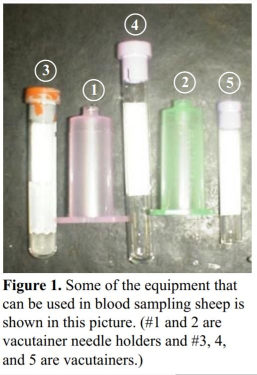
Types of Vacutainers
Certain laboratories require that you ship the blood sample in specific vacutainers. The color of the stopper in the tube and the size of the tube are used for identification of the test being conducted. Purple top tubes used for whole blood shipment are for DNA testing. These tubes contain an anticoagulant that needs to be mixed with the blood once it has been drawn. Red top tubes, or marble top tubes, are used for blood samples that need to clot and be centrifuged, so that the clear serum can be removed and tested. The company should specify the type of tube needed for the laboratory procedure to be done.
Steps to Drawing Blood
1. Shearing
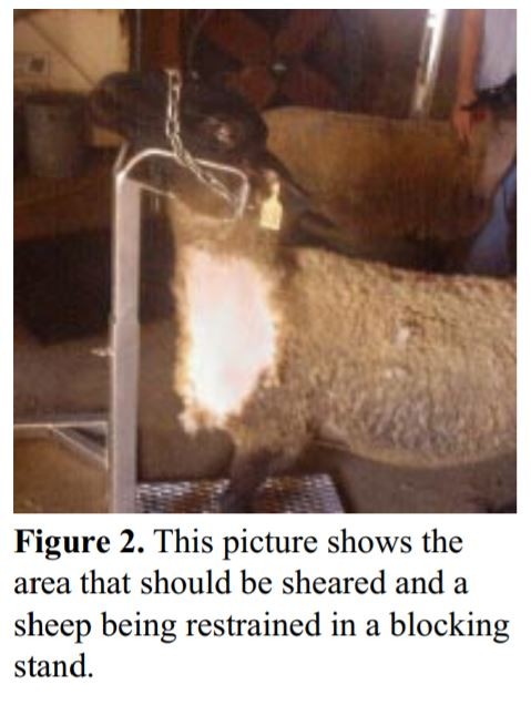
Correctly position the animal for shearing. Use electric shears to shave a patch approximately 4 inches wide by 8 inches long. Figure 2 shows an example of a sheep with a larger patch size for descriptive purposes. Shaving an area allows for easier viewing of the vein and provides a clean area in order to minimize the chance of introducing dirt or bacteria into the vein with the needle.
2. Restraint
Blood sampling can be done with assistance or alone; however, producers who are new at blood sampling will benefit from having assistance. The assistant should turn the head of the animal at a 30-degree angle to the side by holding the animal under its jaw to allow for easy access to the vein. The animal's body may also need to be restrained. Another assistant or a blocking stand (Figure 2) can help keep the animal from moving.
Restraining an animal without assistance is better for those who have become proficient at drawing blood. The handler should straddle the animal, place his or her knees behind the shoulders of the animal, and back the animal in a corner or against a wall to help control its hindquarters. The animal's head should be turned opposite to the side of collection, once again at a 30-degree angle. Restraint of the head is accomplished by using the elbow and the upper arm to keep it held off to the side. This leaves both hands available for the blood collection.
An unruly animal can be dangerous to the assistants, the person drawing the blood, and to itself. It is important to be gentle and patient when restraining the animal.
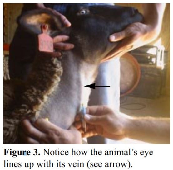
3. Locating the Vein
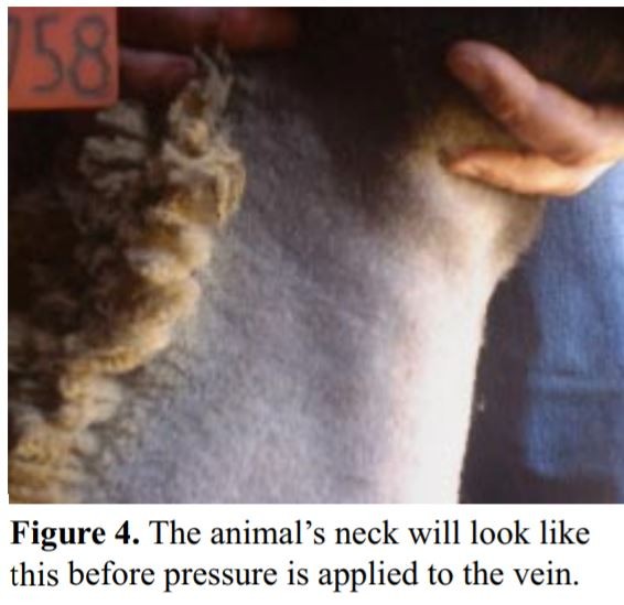
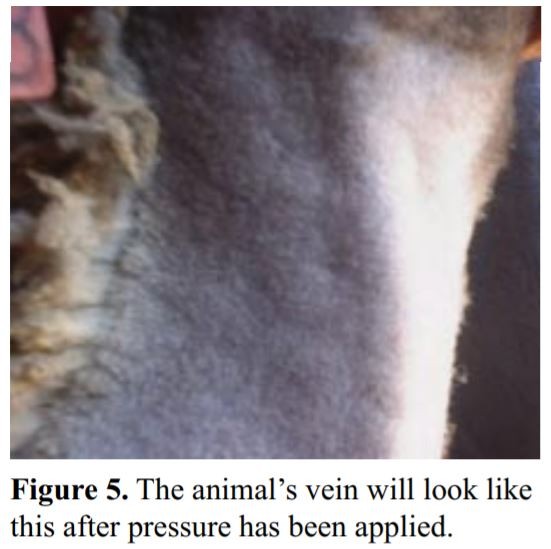
The easiest way to locate the vein is to draw an imaginary line from the middle of the animal's eye down the side of its neck (Figure 3). The vein can be located by applying pressure with the thumb or fingers below the half-way point of the shaved area. The pressure will cause the vein to pop up and be easy to see. The photographs in Figures 4 and 5 illustrate what the vein looks like before and after pressure.
Animals that are overweight may have veins that are harder to locate because of excess fat. If it is difficult to locate the vein, a small amount of alcohol poured over the area where the vein is supposed to be will help locate it.
4. Preparing the Site
Once the vein has been located, the area needs to be properly cleaned to keep bacteria out of the needle insertion site. This is accomplished by using the surgical scrub on the area. Apply a small amount of the surgical scrub to a few pieces of gauze. Squeeze some of the excess scrub out of the gauze before applying it to the patient to make the process easier. The area should be cleaned by starting in the center and working out toward the edge.
Never go back over a place that has already been wiped, because bacteria could be carried back into the clean area.
5. Blood Collection
Once the area has been cleaned and the vein has been located, the blood can be drawn. This can be done by using a needle and syringe method or by using a vacutainer needle and a vacutainer. The first method uses a 6 cc syringe, 20-gauge by one-inch or less needle, and vacutainer.
The other method requires a 20-gauge by one-inch or less vacutainer needle, vacutainer holder, and vacutainer.
Syringe and Needle Method
If a needle and syringe are used, be sure to check that the needle is firmly attached to the syringe and that both the syringe and the needle are new and clean. Contamination from other animals could cause contamination of the sample or infection of the animal.
Remove the cap from the needle first and be careful not to stick yourself. The plunger of the syringe can be easily operated using the thumb, forefinger, and middle finger of the right hand. Before inserting the needle into the vein, pull back on the plunger to break the seal in a new syringe, and then depress the plunger completely to express any air that was drawn into the syringe. Use the other fingers to help guide the needle into the vein at the lowest point possible on the exposed area of the neck. By doing this, the vein can still be used if there are unsuccessful attempts at drawing the blood. Gently pull back on the syringe to see if the needle is in the vein. If no blood pulls back into the syringe, the needle is either parallel to the vein, or it has gone completely through the vein and out the opposite side. Light movements of the syringe can be used to try to locate the vein and penetrate it. When blood is easily pulled back into the syringe, the needle is within the vein. Fill the syringe with the desired amount of blood. Once the sample has been obtained, remove the pressure from the vein, take the needle out, and press gently on the site of needle insertion. Finally, place the needle through the stopper of the appropriate blood collection tube and allow the vacuum in the tube to empty the syringe.
Vacutainer Needle Method
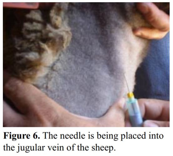
The needle holder should be guided into place with the right hand while the left hand holds off the vein. The vein should be easy to see and feel. Try to aim for the center so you will have more of a chance of placing the needle within the vein.
Before inserting the needle, be sure to check that the flat part of it is to the outside. Inserting the needle does not require a lot of force. Apply enough pressure to break through the skin and enter into the jugular vein (Figure 6).
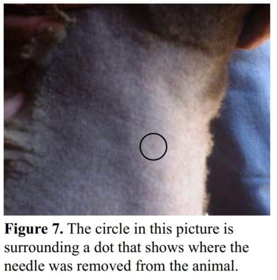
Once the needle is in place, apply pressure so that the vacutainer is pushed up onto the needle. If the needle is in the vein, blood will start to fill the container immediately. If this does not happen, gently move the needle so that the tip comes to the outside of the vein wall and re-insert. Gentle prodding may be needed to achieve maximum blood flow. Before removing the needle, be sure to remove your left hand, which applies the pressure to prevent blood from exiting through the insertion site. Also, before removing the needle, be sure to remove the vacutainer to prevent the loss of the vacuum in the tube. If the needle is removed from the skin, the vacuum will be lost and a new blood collection tube will be needed. After the needle has been removed from the skin, press your fingertip over the area where the needle was inserted. A small red dot may appear on the animal's neck from where the needle was removed (Figure 7). This is normal and is nothing to be concerned about.
Post Collection Handling
The needle and syringe method requires extra work when it comes to processing the sample. The sample should be placed directly from the syringe into the proper vacutainer. Be sure to use an ink pen or a marker to properly label the container with the animal's identification to ensure the test results are accurate. If you used the vacutainer needle method, the blood is already in the proper container for shipment. In either case, the blood should be rocked back and forth a few times to help prevent clotting once it is in the vacutainer. Proper storage of the sample in a refrigerator is important to preserve the sample in purple top tubes. Prompt centrifugation (spinning down) of the blood is important with blood from red or marble top vacutainers. Most blood samples will be sent to outside labs. Samples in the purple top tubes are sent in for DNA testing for Spider Lamb Syndrome and Scrapie Resistance. In the red or marble top tubes, the serum, which is the clear part of the blood, is spun down in a centrifuge after it has clotted. The serum in these tubes is what is tested. All directions should be followed closely for handling and shipping to respective laboratories.
Care of the Patient
Once the blood has been removed from the animal, the insertion site should be swabbed with alcohol to remove any bacteria that might have entered the area during the drawing of the blood. If the vein is still bleeding, apply pressure to the area for ten to fifteen seconds. The pressure causes the blood to clot and the bleeding to cease. If complications while collecting the blood occur, it is possible that a hematoma will appear. If a hematoma does occur, it may remain in place for a day or two before it goes away. The animal should be watched closely for an hour or two to ensure that no bleeding occurs and that the animal exhibits normal behavior.
Clean-up
Throw away the gauze sponges used to clean the injection site in the regular trash. However, the needle (vacutainer or regular) and syringe should be properly disposed of in a Sharps container or set aside for return to the veterinarian for disposal. The needle (vacutainer or regular) and syringe are considered medical waste and need to be disposed of appropriately.
Conclusion
Blood sampling in sheep is not difficult with some experience. Newer technologies have opened up many opportunities for monitoring the health status of sheep. Through persistence and patience, collecting blood samples can become a routine part of flock management. Consulting a veterinarian or another producer who has experience in drawing blood from sheep can help producers learn this management skill.
Blood Sampling in Sheep (pdf; 168KB)

Field Crops
Dairy

Business
Livestock
Grains
Upcoming Events
WNY Pastureland Conversion & Soil Health Field Day
July 16, 2025
Middleport, NY
Join American Farmland Trust for the Western New York Soil Health Field Day on July 16, 2025, at Zeliff Farm in Middleport, NY, from 9:00 AM-3:15 PM. Learn about pasture conversion, soil health benchmarking, biochar in grazing systems, and best grazing practices. Plus, enjoy hands-on demos with the NY Soil Health Trailer, drones, and cover crops! Check out the attached agenda for more information about the field day and REGISTER HERE. Zeliff Farms is a regenerative beef operation who has recently partnered with AFT on outreach and education to farmers including learning circles and evaluating biochar effects on soil health.
IPM Strategies to Protect Corn and Soybean Seed in NY
July 30, 2025
Hamburg , NY
SWNYDLFC and Cornell IPM are hosting a grower meeting to discuss integrated pest management strategies for protecting corn and soybean seed in New York.
FAMACHA Training for Sheep and Goat producers in Woodhull NY
August 13, 2025 : FAMACHA Training in Woodhull
Woodhull, NY
Join us for a discussion and hands-on training for internal parasite integrated pest management in sheep and goats. Certification is available to all students participating in the workshop.
Announcements
No announcements at this time.